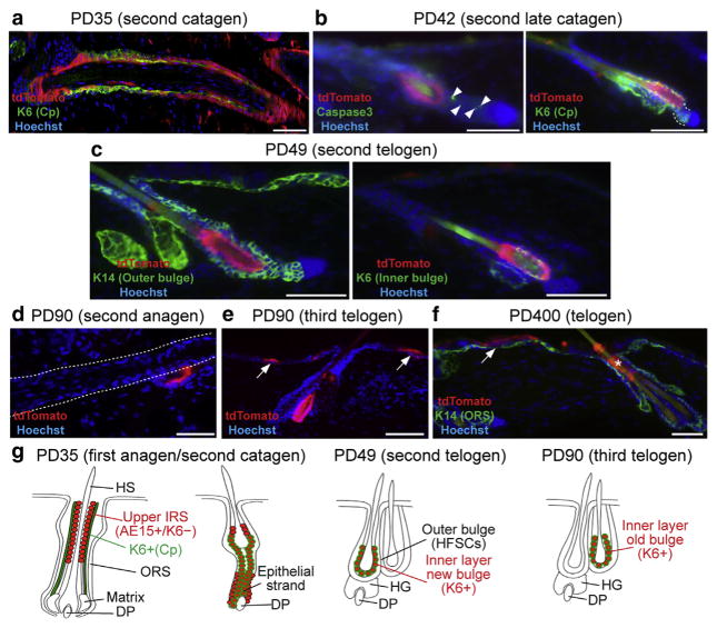
Figure 2. Slc1a3-CreER marked cells are a source for the telogen bulge K6+ inner layer.
(a–f) Images of immunostained lineage traced skin tamoxifen injected as shown in Figure 1 at ages and stages indicated. Confocal images of Z-stacks are shown in Supplementary Figure S3a–d. Arrowheads in (b) represent cell death. The dashed line in (b) represents the lower part of the follicle, where tdTomato+ cells were not colocalized with K6. The dashed line in (d) bounds the anagen follicle. Note localization of tdTomato+ cells in the old bulge in this image. The asterisk in (f) shows remnants of tdTomato+ cells shed through the infundibulum. Arrows represent tdTomato+ clones in the interfollicular epidermis. Scale bars = 50 μm. (g) Summary cartoon with tdTomato in red and K6 in green. Abbreviations are the same as in the Figure 1 legend.

