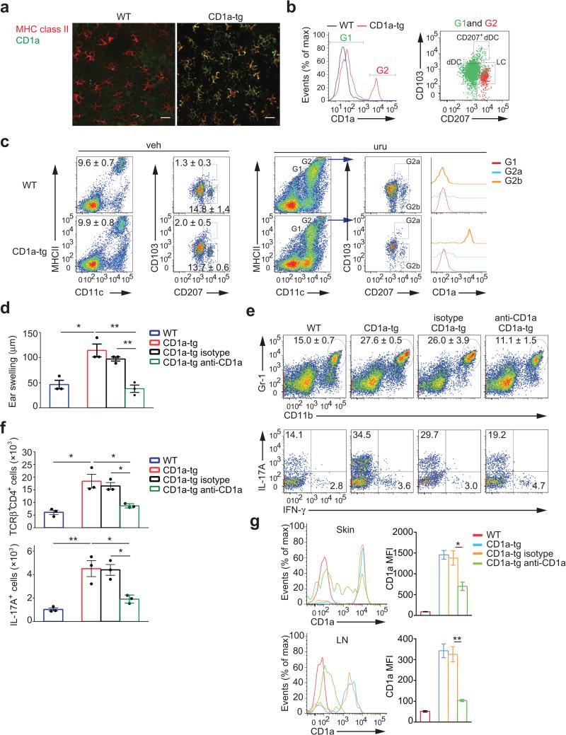
Figure 4.
CD1a expression on Langerhans cells is essential for generation of TH17 cells and dermatitis. (a) Confocal microscopy of epidermal sheets stained for MHC class II and CD1a. Scale bar: 20 μm. (b) Flow cytometry of CD1a expression on CD11c+MHCIIhi cells (left panel), CD103 and CD207 (Langerin) expression in CD1a-negative (I, green dots) and CD1a-positive (II, red dots) populations of CD1a-tg mice gated from the left plot (right panel). (c) Flow cytometry of Langerhans cells population in ear cells isolated from mice (n = 3 per group) sensitized to urushiol on day 0 and challenged with vehicle (veh) or urushiol (uru) on day 5, assessed on day 2 after challenge. CD1a expression on CD11c+MHCIIint (Gate, G1), CD103+CD207+ (G2a), and CD103−CD207+ (G2b) cells among CD11c+MHCIIhi (G2) cells of urushiol-challenged mice was further analyzed (far right). (d-g) Ear swelling (d), frequencies of Gr-1hiCD11bhi granulocytes (e, top), IL-17A+ or IFN-γ+ cells among CD45+TCRγ+CD4+cells (e, bottom), absolute cell numbers of CD4+ T cells (f, top), IL-17A-producing CD4+ T cells (f, bottom), and CD1a expression on LCs in ear skin (g, top) and dLN (g, bottom) from urushiol-challenged wild-type, CD1a-tg, or CD1a-tg mice injected with anti-CD1a or isotype (n = 3 per group). MFI indicate mean fluorescent intensity. Each symbol represents an individual mouse (d,f). Data shown are the mean ± s.e.m. * P < 0.05, ** P < 0.01; NS, not significant, using one-way ANOVA and multiple comparisons. Data are representative of four independent experiments with similar results.

