Fig. 1.

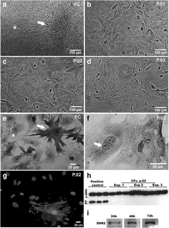
Characterization of culture enriched in cardiac fibroblast. a-f Phase contrast and bright field microscopy showing features of CFs cultures. a, e Primary culture presenting cardiomyocytes clusters (arrow) that showed spontaneous contraction, surrounded by CFs (*) forming a monolayer. b-d Passages 1, 2 and 3, respectively, showing the aspect of CF-enriched cultures. f Fibroblast culture at passage 2, stained with Giemsa, demonstrating typical morphology with elongated cells, cytoplasmic extensions, the oval and large nucleus with apparent nucleoli. g Immunofluorescence showing sarcomeric tropomyosin expression indicating 95% purity of CFs (nuclei were labeled with DAPI). h Immunoblotting for ST revealed that CF cultures were myocyte-free. For positive controls, hearts of mouse embryos were used. Lanes show technical triplicates of three independent experiments (EXP). α-SMA expression, used as load control, can be observed in all samples. i Representative immunoblotting demonstrating DDR2 expression in CFs in different times of culture
