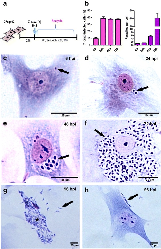
Fig. 2.

Trypanosoma cruzi intracellular cycle in cardiac fibroblasts. a Schematics of the experimental design: fibroblast cultures were infected at passage 2 and analyzed after 6 to 96 h of infection (MOI 10). b Quantitative data of the infection of CF by T. cruzi. At 6 hpi 9.4% of the host cells are infected by one parasite and from 24 to 72 hpi the infectivity was of 38%. Proliferation of the intracellular amastigotes started at 48 hpi with 5 parasites/infected cell and reached 40 parasites/infected cell at 72 hpi. c-h Bright field microscopy representative images of T. cruzi infected CFs cultures stained with Giemsa showing the parasite intracellular cycle. In c intracellular parasites are visible already after 6 h of infection (arrow). d At 24 h post-infection we can observe the beginning of the amastigotes proliferation process (arrow). e At 48 h an increased number of intracellular amastigotes can be observed. f After 72 h of infection a high number of amastigote forms are seen through all cytoplasm. g At 96 h of infection is possible to see that the parasites differentiated to trypomastigotes (asterisk) and evaded the host cells (arrow). h After evading host cells, the released parasites attach to new CFs to restart a new cycle (arrow)
