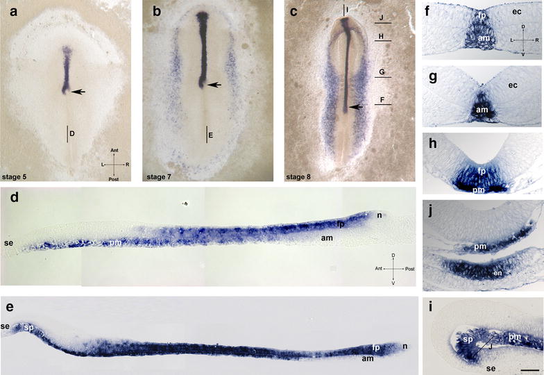
Fig. 1.

Temporal dynamics of shh expression in the chick. a–c whole-mount views of embryos at HH stage 5 (a), 7 (b) and 8 (c); median sagittal technovit sections of stage 5 (d) and stage 7 (e); f–j transversal sections of HH stage 8 embryos at the levels shown in c. i median sagittal section of stage 8 embryo. Labeling: fp floor plate, am axial mesoderm, ec ectoderm, en endoderm, n node, pm prechordal mesoderm, sp preoral gut (prospective area of Seessel’s pouch), se superficial ectoderm, arrow—position of the node. Intersecting arrows indicate anatomical axes: A anterior, P posterior, L left, R right, D dorsal, V ventral. Scale bar 400 µm (a–c), 75 μm (e) and 50 µm (d, f–i)
