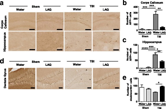
Fig. 2.

Laquinimod has neuroprotective role on TBI. a Representative × 20 images of APP staining in the corpus callosum and hippocampus on ipsilateral side of injury (or craniotomy for sham) from each group. b–c Quantification of the APP-positive cells reveals tendency of decreased axonal damage both in corpus callosum and in hippocampus in the TBI-laquinimod group compared to the TBI-water group (n = 3 mice for each sham group, n = 4 mice for each TBI group). d Representative × 20 images of doublecortin staining in the dentate gyrus on ipsilateral side of injury (or craniotomy for sham) from each group. e Quantification of neurogenesis in dentate gyrus reveals a significant increase in TBI-laquinimod group compared to the TBI-water group. Symbols denote significant differences between groups. Error bars indicate mean ± s.e.m. (n = 5 mice for each group). *p < 0.05, **p < 0.01, ***p < 0.001
