
Fig. 5.
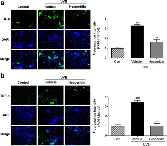
Images of immunohistochemically-stained skin tissue. Immunofluorescence study was carried out using (a) IL-8 and (b) TNF-α antibodies. Blue staining represents nucleus. Quantitative analysis of fluorescence intensity was performed by Image J software. ##P < 0.01, and ###P < 0.001 significant difference from control and **P < 0.01, and ***P < 0.001 Significant difference from UVB/vehicle groups