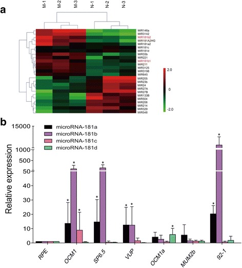
Fig. 3.

The expression profile of miR-181 in melanoma tissues and UM cells. a Hierarchical clustering analysis of miRNAs that were differentially expressed in melanoma compared with non-tumor samples. Expression values are represented in shades of red and green indicating expression above and below the median expression value across all samples (log scale 2, from − 2 to + 2), respectively. miR-181b1 and miR-181b2 were significantly upregulated in melanoma tissues. b The expression of miRNA-181a-d was measured by qRT-PCR in RPE, OCM1, SP6.5, VUP, OCM1a, MUM2b and 92-1 cells. miR-181b was overexpressed in OCM1, SP6.5, VUP and 92-1 cells by approximately 50-fold and more than 1000-fold in 92-1 cells, while miR-181b was not upregulated in OCM1a or MUM2b cells. miR-181a was upregulated in OCM1, SP6.5, VUP and 92-1 cells by approximately 12-to-20-fold. The expression levels of miR-181c and miR-181d were not upregulated in most UM cell lines, except for a slight increase in miR-181c in OCM1 cells and miR-181d in OCM1a cells, both less than 10-fold. There was no downregulation of any miR-181 family members. Triplicate assays were performed for each sample, and the relative level of each miRNA was normalized to U6 (*P < 0.05)
