Fig. 5.
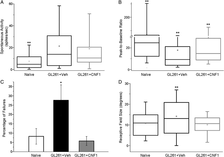
Sparing of functional properties of cortical units by CNF1 treatment. (A) Spontaneous firing of neurons in naïve (n = 11, 164 cells recorded), vehicle-injected (Veh; n = 8, 121 cells recorded), and CNF1-injected (n = 9, 111 cells recorded) glioma-bearing mice. Both glioma groups differ from naïve (ANOVA on ranks, post hoc Dunn's test, **P < .01). (B) Neuronal responsivity (peak firing evoked by visual stimulation divided by spontaneous activity) in naïve, vehicle, and CNF1 glioma-bearing mice. Compared with naïve mice, glioma-bearing animals display a lower responsivity which is partially counteracted by CNF1 (1-way ANOVA on ranks, post hoc Dunn's test, glioma-bearing vehicle vs CNF1, **P < .01). (C) Percentage of failures (lack of response to a light bar drifting into the receptive field) of cortical units. Note the higher percentage of failures in vehicle animals with glioma (1-way ANOVA, post hoc Holm–Sidak test, *P < .05). (D) Box charts showing neuronal receptive field size in naïve animals, and vehicle- and CNF1-treated glioma-bearing mice. Note the increase in receptive field size for vehicle glioma-bearing mice compared with naïve and CNF1-treated group (1-way ANOVA on ranks, post hoc Dunn's test, **P < .01).

