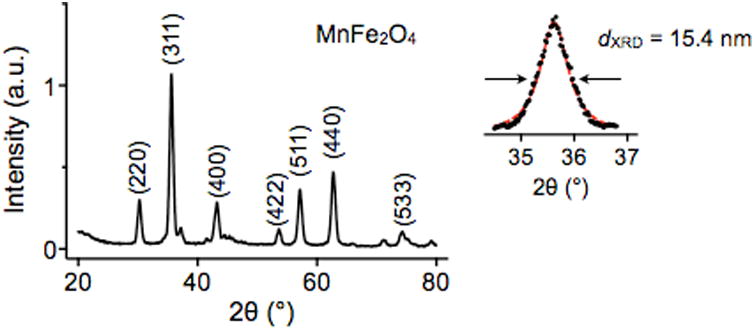
Figure 14. Structural analysis by X-ray diffraction (XRD).

By scanning the incident angle (θ) of an X-ray beam and using powdered MNPs, the diffraction peaks of different lattice planes are measured. A diffraction pattern from powdered MnFe2O4 MNPs is shown, which confirms to that of a typical spinel structure of ferrite. The crystal size is also estimated by fitting the major peaks (311) to Scherrer's formula. Reproduced with permission from Ref. 44. Copyright 2009 National Academy of Sciences, USA.
