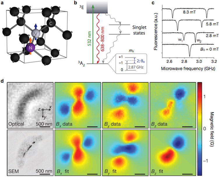
Figure 6. Diamond-based magnetometer.

(a) Structure of nitrogen (N) and vacancy (V) inside a diamond lattice. C, carbon. The blue and orange arrows indicate the electron and nitrogen nuclear spins, respectively. (b) Energy state diagram. The N-V center has a spin-triplet ground state (3A2) with a 2.87 GHz zero-field splitting between the ms = 0 and ms = ±1 spin states. Optical excitation (532 nm) produces the excitation state (3E) which decays back to the ground state by emitting a photon (638–800 nm wavelength). The ms = 0 spin state has a stronger fluorescence than the ms = ±1 states, because the ms = ±1 excited states also decay non-radiatively via metastable singlet states. When an external field (B0) is applied, the ms = ±1 states are split by 2γB0, where γ is the gyromagnetic ratio of the N-V electronic spin. (c) Optically detected magnetic resonance spectra for a single nitrogen-vacancy. The splitting between w1 and w2 is linearly proportional to B0. (d) Detection of magnetotactic bacteria with a N-V diamond sensor. Left top and bottom images are from optical and scanning electron microscopy (SEM), respectively. Measured magnetic field projections of the bacterium along the x (Bx), y (By) and z axes (Bz) are shown in the top row. The bottom row shows simulated magnetic field projections, assuming that MNP locations match those in the SEM image. Reproduced with permission from Refs. 73, 70, and 75. Copyright 2008, 2012, 2013 Nature Publishing Group.
