
Figure 1. Effect of exogenous insulin implant-induced hyperinsulinemia on atherosclerosis and its complexity in the ApoE−/− mice fed on HFD.
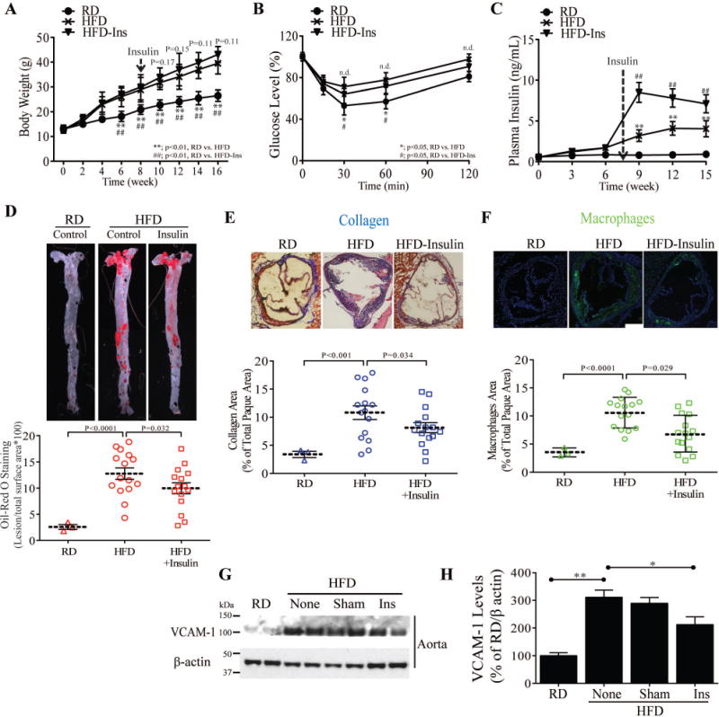
(A) Body weights of experimental animals during HFD feeding (n = 9 for control, n = 9 for insulin treatment). (B) Insulin sensitivity (IPITT) (C) Time course of plasma fasting insulin. (D) en face staining and quantification of aortas as percent of lesion area without or with insulin treatment (n = 10 per group). (E and F) Representative examples and quantification of cross-sections from the aortic sinus stained with trichrome (E) and MAC2(F). (G and H) Immunoblot and densitometry of VCAM-1 levels in aorta from control, insulin treated and phlorizin-treated ApoE−/− mice fed on HFD. All data are presented as mean ± SEM. *p < 0.05, **p < 0.01. P-values for panels A, B, and C were computed using linear mixed effects models to account for the repeated measures within animals over time. P-values for panels A, B, and C were computed using linear mixed effects models to account for the repeated measures within animals over time. P-values for panels D, E, F, and H were computed using general linear model (ANOVA). In all cases, post hoc t-tests with Tukey’s adjustment for multiple comparisons were conducted where overall F-tests were significant to ascertain the location of any significant pairwise differences.