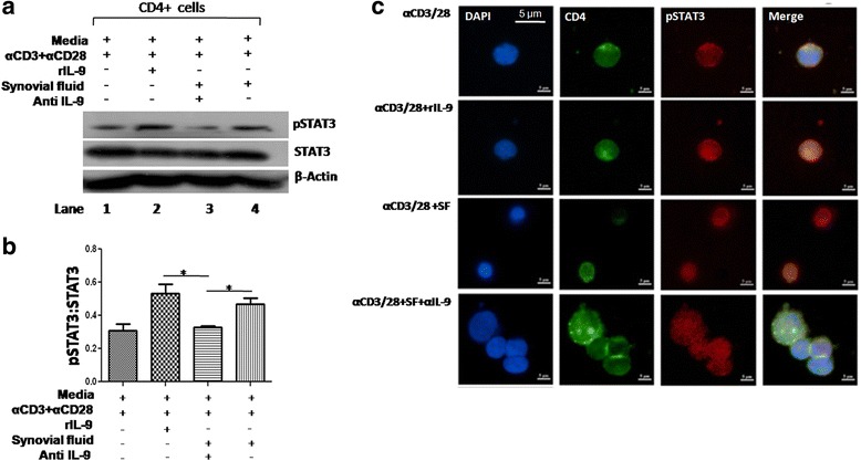
Fig. 6.

IL-9 induces transcription factor for Th17 cells. a Representative western blot image shows phosphorylation of STAT3 (pSTAT3) in the presence of rIL-9 and SF. pSTAT3 was enhanced with rIL-9 and SF (lane 2 and 4) and in presence of anti-IL-9 antibody in SF reduced pSTAT3 (lane 3, n = 2). b Bar graph shows cumulative densitometry of pSTAT3 (pSTAT3:STAT3, β-actin as loading control, n = 5, unpaired t test, mean ± SEM, *p < 0.05).c Representative confocal micrograph from two independent experiments, shows phosphorylation of pSTAT3 (red), nucleus (DAPI, blue) in CD4+ T cells (green) and nuclear translocation (merged image in violet). IL-9R interleukin 9 receptor, SF synovial fluid, STAT3 signal transducer and activator of transcription 3, α IL-9 anti-interleukin 9
