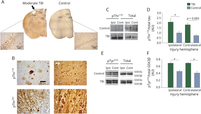
Figure 4. Tau phosphorylated at threonine 175 (pThr175 tau) pathology in moderate traumatic brain injury (TBI).
At 3 months following moderate TBI, pThr175 tau pathology was observed. (A) Composite images of whole brain sections stained for pThr175 tau. Images were taken with a 4× objective. Inlay image taken with 40× objective. Arrow denotes site of injury. CA1 = CA1 region; CA3 = CA3 region; DG = dentate gyrus. Scale bar = 50 μm. (B) High-magnification images with moderate TBI show neuronal and neuritic pathology. Images were taken with 100× objective. Scale bar = 20 μm. (C) Western blots of pThr175 tau and total tau in ipsilateral and contralateral brain injuries. (D) Densitometry of western blots probed for pThr175 tau and total tau (pThr175/total tau). (E) pTyr216 glycogen synthase kinase–3β (GSK3β) and total GSK3β (pTyr216/total GSK3β) in ipsilateral and contralateral brain injuries. (F) Densitometry of western blots probed for pTyr216 GSK3β and total GSK3β. *p < 0.05. Contra = contralateral injury hemisphere; Ipsi = ipsilateral injury hemisphere.

