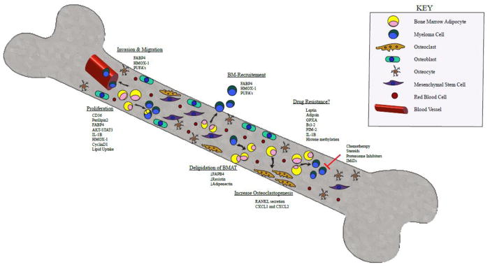
Figure 1. Multistep Signaling between Bone Marrow Adipocytes and Multiple Myeloma.
A schematic overview of the signals sent and received between BMAs and myeloma cells within the bone marrow microenvironment. MAT may promote MM progression, and MM cells may lead to MAT destruction. Signaling from MAT through FABP4, HMOX-1, and PUFAs promotes MM invasion and migration through blood vessel walls into the bone marrow. In addition, the same signals aid in MM recruitment to the bone marrow. MAT can also induce MM cell proliferation through multiple mediators such as CD36, perilipin2, FABP4, AKT-STAT3, IL-1β, HMOX-1, CyclinD1 and direct uptake of lipids. We hypothesize that MAT may play a role in MM drug resistance through mechanisms involving leptin, adipisin, GPXA, Bcl-2, PIM-2, IL-1β and modification of histone methylation. Not only can MAT influence MM cells, it has been shown to increase osteoclastogenesis through secretion of RANKL and CXCL1/CXCL2. Lastly, MM cells can induce MAT delipidation potentially by downregulating FABP4, adiponectin and resistin.
Abbreviations: BMA: Bone Marrow Adipocyte. FABP4: fatty acid binding protein 4; HMOX-1: Heme Oxygenase 1; PUFAs: polyunsaturated fatty acids; CD36: platelet glycoprotein 4; IL-1B: Interleukin 1 Beta; GPXA: glutathione peroxidase related gene; Bcl-2: B-cell lymphoma 2; PIM-2: Proviral Integrations of Moloney virus 2; RANKL: Receptor activator of nuclear factor kappa-B ligand; CXCL1/CXCL2: chemokine ligand 1 and 2.

