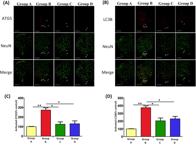
Fig 5. The Effects of EPO on autophagy markers in burn injury model by immunofluorescence analysis.
(A, B) Double immunofluorescence staining and merged images using LC3B, ATG5, and NeuN in the spinal cord ventral horn were shown. The quantitative analysis of ATG5 (C) and LC3B activity (D) were measured. Burn injury significantly increased LC3B and ATG5 immunoreactivity in Group B versus Group A. After EPO treatment, LC3B and ATG5 immunoreactivity decreased markedly in Groups C and D vs Group B (**: p < 0.01; *: p < 0.05 versus Group B; Group A: 100%). Scale bars: 50 μm.

