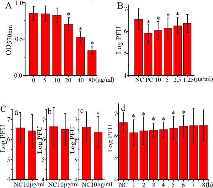
Fig 1. Anti-IAV activity of rhein in vitro.
(A) The cytotoxicity of rhein was determined by a MTT method, *P < 0.05 vs. the blank control (0 μg/mL). (B) Inhibition of rhein on IAV (ST169, H1N1) replication was determined by a plaque inhibition assay. In the negative control (NC), MDCK cells were infected with IAV (MOI = 0.001) but not treated with any drugs; in the positive control (PC) and rhein-treated groups, MDCK cells were infected (MOI = 0.001) and treated with ribavirin (25 μg/mL) and rhein (1.25, 2.5, 5, and 10 μg/mL), respectively. After 48 h p.i., the supernatants were harvested and the titers were determined by a plaque formation assay. (C) The results of the time-of-addition assay, which contained four tests: (a) direct inactivation assay, (b) influence-on-cell assay, (c) influence-on-viral adsorption assay, and (d) different-time-points p.i. assay. MOI = 2.0. 0.5% DMSO was used as the negative control (NC). After 12 h p.i., the supernatants were harvested and the viral titer was determined by a plaque formation assay. The experiment was repeated five times and each experimental condition was performed in triplicate (n = 5). All data shown were mean ± SD. *P < 0.05 vs. the NC group.

