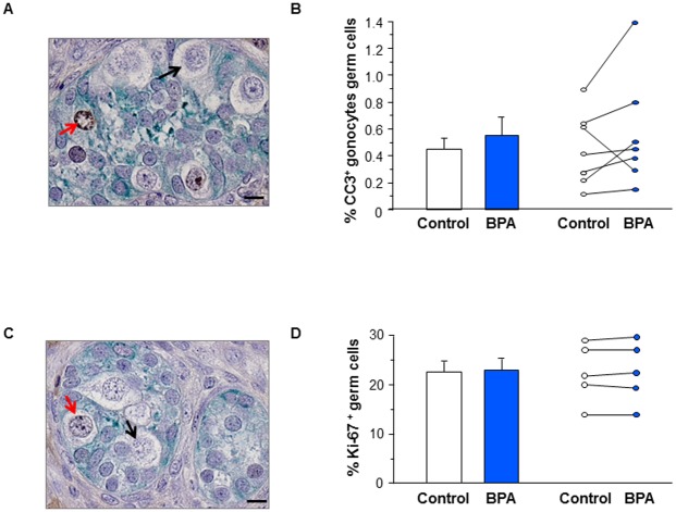
Fig 4. Effect of BPA exposure on germ cell apoptosis and proliferation in first trimester human fetal testis xenografts.
Human fetal testes (9.1–11.3 GW) were xenografted into castrate Nude (host) mice. Host mice received vehicle (Control) or 10μM BPA in the drinking water for five weeks. (A) Histological sections of the testis after labeling with anti-CC3 antibody (brown) and anti-AMH antibody (green). (B) Quantification of CC3+ cells displayed as mean ± SEM (n = 7) on the left panel and as individual values with a line drawn between the control and the corresponding BPA-treated testis from the same fetus on the right panel. (C) Histological sections after labeling with anti-Ki-67 antibody (brown) and anti-AMH antibody (green). Positive (red arrows) and negative (black arrows) germ cells can be identified. (D) Quantification of Ki67+ gonocytes displayed as mean ± SEM (n = 5) on the left panel and as individual values with a line drawn between the control and the corresponding BPA-treated testis from the same fetus on the right panel. Scale bars: 15 μm. Data analyzed using Wilcoxon paired test. For both apoptosis and proliferation, the differences between vehicle and BPA-exposed groups were not statistically significant.

