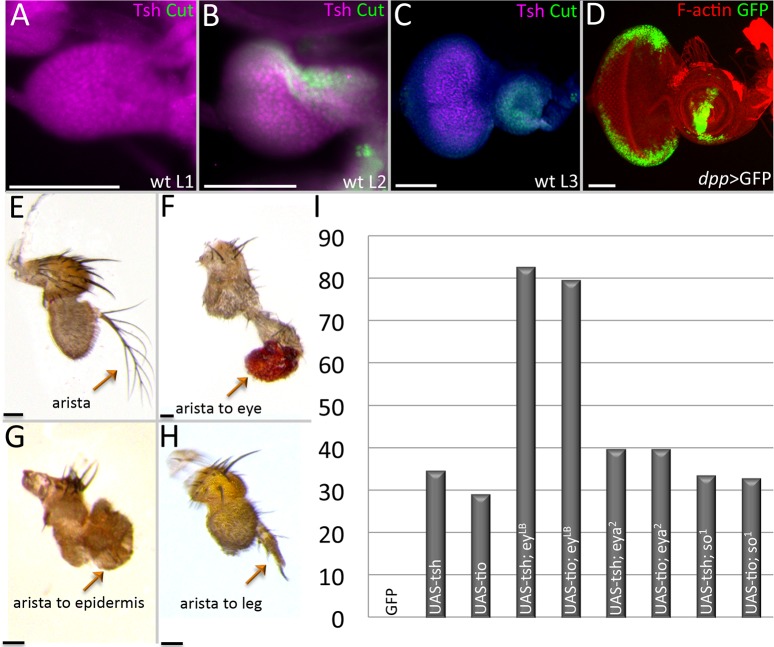
Fig 7. Continued expression of tsh/tio in the antennal disc alters its fate.
(A-D) Light microscope images of eye-antennal discs. (A) Wild type first instar eye-antennal disc. Tsh protein is expressed throughout the entire disc. Cut protein is not yet detected. (B) Wild type second instar eye-antennal disc. Tsh protein continues to be found throughout the disc while Cut protein is found within the antennal portion. (C) Wild type third instar eye-antennal disc. Tsh protein is found just within the undifferentiated cells of the eye field while Cut protein is exclusively seen in the antennal field. (D) dpp-GAL4, UAS-GFP. The dpp-GAL4 construct drives expression along the posterior-lateral margins of the eye field and within a ventral sector of the antennal disc. (E-H) Light microscope images of adult antennal segments. (E) Wild type antenna with arista. (F) dpp-GAL4, UAS-tsh; the arista has been converted into an ectopic eye. (G) eya,2 dpp-GAL4, UAS-tsh; the arista is transformed into head epidermal mass. (H) dpp-GAL4, UAS-tsh, eyLB; the arista has been transformed into the leg tarsal segment. (I) Chart documenting the percentage of arista to leg transformations when tsh/tio are expressed in wild type and RD gene mutants (N ≥ 35).(Scale bars, 50 μm).

