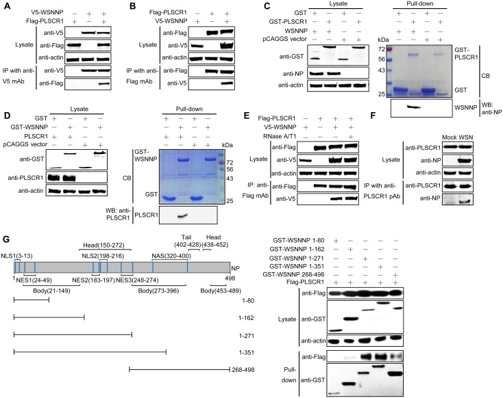
Fig 2. NP interacts with PLSCR1 in mammalian cells.
(A, B) Co-IP assay of V5-NP and Flag-PLSCR1 in HEK293T cells. HEK293T cells were transfected individually or in combination with plasmids expressing V5-WSNNP and Flag-PLSCR1. Cell lysates were immunoprecipitated with a mouse anti-V5 mAb (A) or a mouse anti-Flag mAb (B) and were subjected to western blotting with a rabbit anti-V5 pAb or a rabbit anti-Flag pAb to reveal the presence of NP and PLSCR1, respectively. (C, D) GST pull-down assay of NP and PLSCR1. Lysates of HEK293T cells transfected with the GST or GST-PLSCR1 construct were incubated with Glutathione Sepharose 4 Fast Flow and then mixed with lysates from cells transfected with pCAGGS or pCAGGS-WSNNP (C); HEK293T cell lysates containing exogenously expressed GST or GST-WSNNP were incubated with Glutathione Sepharose 4 Fast Flow and then mixed with lysates from cells transfected with pCAGGS or pCAGGS-PLSCR1 (D). After washing away the unbound proteins, equal volumes of proteins bound to the beads and the original cell lysates (5% of the input) were examined by western blotting using a rabbit anti-NP pAb, a rabbit anti-GST pAb, or a rabbit anti-PLSCR1 pAb. GST, GST-PLSCR1, or GST-WSNNP proteins in the eluates were detected by Coomassie blue (CB) staining. (E) The NP-PLSCR1 interaction does not rely on RNA binding. HEK293T cells were transfected individually or in combination with plasmids expressing V5-WSNNP and Flag-PLSCR1. Cell lysates treated with RNase A/T1 or left untreated were immunoprecipitated with a mouse anti-Flag mAb and were subjected to western blotting with a rabbit anti-V5 pAb or a rabbit anti-Flag pAb to reveal the presence of NP and PLSCR1, respectively. (F) PLSCR1 interacts with NP during natural viral infection. Confluent A549 cells grown in 6-well plates were mock infected with PBS or infected with WSN virus at an MOI of 5. At 6 h p.i., cell lysates were immunoprecipitated with a rabbit anti-PLSCR1 pAb and were subjected to western blotting with a mouse anti-NP mAb or a rabbit anti-PLSCR1 pAb to detect NP and PLSCR1, respectively. (G) Mapping of the PLSCR1-interacting domain within NP. Schematic presentation of influenza NP showing the different domains as well as the truncation mutants made in this study is on the left side. The interaction between PLSCR1 and the NP truncation mutants is shown on the right side. Lysates of HEK293T cells were pulled down with glutathione magnetic beads. The bound proteins were subjected to western blotting with a rabbit anti-Flag pAb or a rabbit anti-GST pAb to reveal the presence of PLSCR1 and NP, respectively. NES, nuclear export signal; NAS, nuclear accumulation signal.

