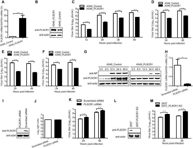
Fig 3. PLSCR1 negatively regulates influenza virus replication.
(A, B) Establishment of an A549 cell line stably overexpressing PLSCR1. The stable overexpression of PLSCR1 was confirmed by quantitative reverse-transcription PCR (RT-qPCR) (A) and western blotting with a rabbit anti-PLSCR1 pAb (B) in comparison with the A549 control cell line transduced with an empty retrovirus. **, P < 0.01. (C, D, E, F) Virus replication in PLSCR1-overexpressing A549 cells. The PLSCR1-overexpressing or empty retrovirus-transduced control A549 cells were infected with WSN (H1N1) (C), AH05 (H5N1) (D), AH13 (H7N9) (E) or FZ09 (H1N1) (F) at an MOI of 0.1. Supernatants were collected at the indicated timepoints, and virus titers were determined by means of plaque assays on MDCK cells. **, P < 0.01. (G) Expression of PLSCR1 and NP in virus-infected cells. The PLSCR1-overexpressing or empty retrovirus-transduced control A549 cells were infected with WSN virus at an MOI of 0.1. Whole cell lysates were collected at the indicated timepoints and subjected to western blotting with a rabbit anti-PLSCR1 pAb or a rabbit anti-NP pAb. (H, I) siRNA knockdown of PLSCR1 in A549 cells. A549 cells were transfected with siRNA targeting PLSCR1 or with scrambled siRNA for 48 h. Whole cell lysates were then collected and analyzed by RT-qPCR (H) or western blotting with a rabbit anti-PLSCR1 pAb (I). *, P < 0.05. (J) Cell viability of siRNA-treated A549 cells was measured by using a CellTiter-Glo assay. The data are presented as means ± standard deviations (SD) for triplicate transfections. (K) Virus replication in siRNA-treated A549 cells. Cells transfected with siRNA were infected with WSN virus at an MOI of 0.1. Supernatants were collected at 24 and 48 h p.i. and titrated for infectious virus by means of plaque assays on MDCK cells. **, P < 0.01. (L) Generation of PLSCR1-KO HEK293T cells. PLSCR1-KO cells were generated by using the CRISPR/Cas9 system. PLSCR1 knockout was confirmed by western blotting with a rabbit anti-PLSCR1 pAb. (M) Virus replication in PLSCR1-KO HEK293T cells. PLSCR1-KO HEK293T or control cells were infected with WSN virus at an MOI of 0.1. Supernatants were collected at 24 and 48 h p.i., and virus titers were determined by means of plaque assays on MDCK cells. **, P < 0.01.

