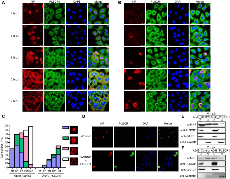
Fig 4. PLSCR1 inhibits the nuclear import of NP.
(A, B) Empty retrovirus-transduced control A549 cells (A) or PLSCR1-overexpressing A549 cells (B) were infected with WSN virus at an MOI of 5. At 4, 6, 8, 10, and 12 h p.i., the infected cells were fixed and stained with mouse anti-NP mAb and rabbit anti-PLSCR1 pAb, followed by incubation with Alexa Fluor 488 donkey anti-rabbit IgG (H+L) (green) and Alexa Fluor 633 goat anti-mouse IgG (H+L) (red). The nuclei were stained with DAPI. (C) Quantitative analysis of NP localization in virus-infected cells. On the basis of the confocal microscopy in panels A and B, the localization of NP (indicative of vRNP) after nuclear import was categorized into four types, clear nuclear localization, simultaneous localization at the edge of the nucleus and the cytoplasm, predominant cytoplasmic localization, and close to the cytoplasmic membrane. The results shown are calculated from one hundred cells viewed under a confocal microscope with a 40X objective lens. (D). PLSCR1 inhibits the nuclear import of NP in transfected A549 cells. A549 cells were transfected with pCAGGS-WSNNP alone or were cotransfected with pCAGGS-WSNNP and pCAGGS-PLSCR1 and were assessed by confocal microscopy. NP was detected with a mouse anti-NP mAb and visualized with Alexa Fluor 633 (red). PLSCR1 was detected with a rabbit anti-PLSCR1 pAb and visualized with Alexa Fluor 488 (green). Yellow in the merged image indicates the colocalization of NP and PLSCR1. (E) PLSCR1-overexpressing or empty retrovirus-transduced control A549 cells were infected with WSN virus at an MOI of 5. At 6 h p.i., the cells were separated into nuclear (N) and cytoplasmic fractions (C). Each fraction was subjected to western blotting with a rabbit anti-NP pAb and a rabbit anti-PLSCR1 pAb for protein detection. (F) PLSCR1-overexpressing A549 cells or control A549 cells were treated with CHX to inhibit protein synthesis. The treated cells were infected with WSN virus at an MOI of 5, and were separated into nuclear and cytoplasmic fractions at 2 h p.i., followed by western blotting to detect the amount of NP in the nuclear and cytoplasmic fractions.

