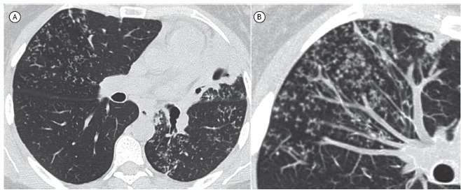
Figure 1. In A, axial CT scan of the chest with lung window settings at the level of the subcarinal region, showing left lung volume reduction and consolidation with cavitation in the lingula. Note also centrilobular branching opacities (tree-in-bud pattern) in the middle lobe and the left lower lobe. In B, image enhancement (maximum intensity projection) demonstrating the tree-in-bud pattern.

