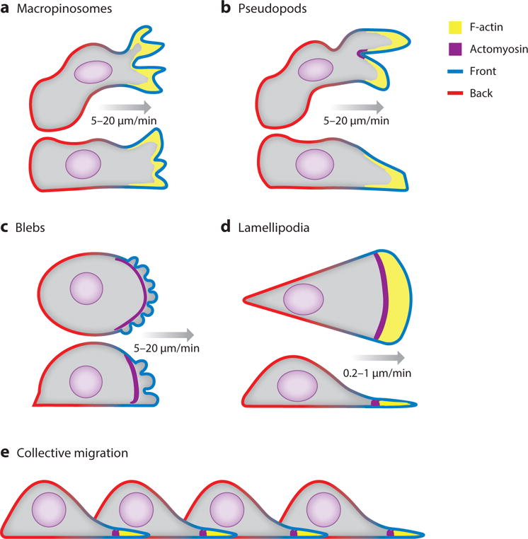
Figure 1.

The diverse array of migratory cell projections that project the cell forward are diagrammed in coronal and sagittal slice. Green membrane represents the “front” and red represents the “back” In these cartoons, only the polymerizing F-actin and contractile actomyosin at the leading edge of cells is highlighted. A) Macropinosomes are wide cup-like shaped structures at the top and sides of the cell. B) Pseudopods are narrower protrusions usually found closer to the substrate. C) Blebs are a result of the plasma membrane detaching from the actomyosin cortex due to contractile pressure. D) Lamellipodia are sheet like structures containing distinct actin and actomyosin zones. E) Collective migration of cells connected and partially driven by cryptic lamellipodia.
