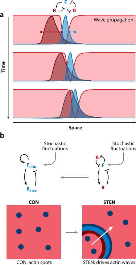
Figure 5.

A. A cartoon of how an excitable wave propagates on the membrane. The reciprocal “front” (F) and “back” (B) zones (green and red, respectively; representative activities as in Figure 4), propagate and the “refractory” (R) zone (deep red) trails, ensuring unidirectional front propagation. B. Coupling between cytoskeletal (CON) and signaling (STEN) activities generate propagating waves of cytoskeletal activity. In the absence of STEN activity, there are flashes of actin polymerization - shown by the blue spots in the box on the left (a random 2D area on the cortex). Once STEN activity is triggered, it causes the CON spots to expand in space – driven by the excitable wave front (green) – creating actin waves.
