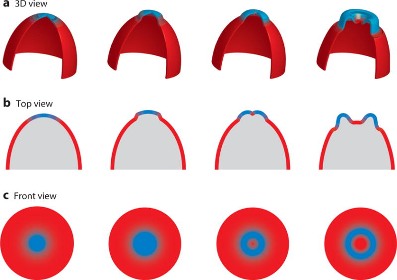
Figure 6.

Cartoon showing the coupling between wave propagation and topology of cellular protrusions. (Top) Three-dimensional representation of the formation of cup-like structures on the cortex as a wave propagates (green and red as “front” (F) and “back” (B) activity, respectively). (Middle and Bottom) A top and front view of these structures shows how these protrusions are born out of wave propagation as the front activity spreading outward creates a hole in 2D, which translates to a cup-like protrusion in 3D.
