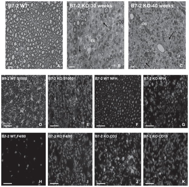
Figure 3.
Pathological characterization of sciatic nerves in SAPP. Toluidine blue-stained photomicrographs of representative 1 μm semi-thin axial sections of plastic-embedded sciatic nerves at different ages are shown (A–C). There is normal axonal density with large, medium, and small myelinated axons in B7-2 WT mice at all ages (A), with rare endoneurial leukocytes seen. Increased leukocyte infiltration (red arrows) with uniformly persistent demyelination (black arrows) and endoneurial edema (EE) are seen in the sciatic nerves at peak severity, as observed at 30 weeks of age in a severely affected B7-2 KO mouse (B). During the late stages of SAPP (C), there is onion-bulb formation (OB), persistence of thinly myelinated and demyelinated axons (black arrows) and foci of infiltrated leukocytes (red arrow), as commonly observed in CIDP nerve biopsies. Digitally merged photomicrographs of representative indirect immunohistochemical 10 μm axial cryostat sections of sciatic nerves stained to detect S100β (Schwann cell/myelin marker: D and E), neurofilament heavy chain (NF-H; axon marker: F and G), F4/80 (macrophage marker: H and I), CD3 (T-cell receptor: J) and CD19 (B-cell marker; K) in B7-2 WT and KO NOD mice at 40 weeks of age are shown. The normal honeycomb appearance of myelinated axons (green immunoreactivity) with scattered nuclei (DAPI stain: blue immunoreactivity) seen in B7-2 WT mice (D) is disrupted, with increased leukocyte infiltration (increased blue immunoreactivity) seen in a SAPP-affected B7-2 KO mouse (E). Normal axonal density (red immunoreactivity) in the same B7-2 WT mouse is seen (F), in contrast to significant axonal loss associated with leukocyte infiltration in the SAPP-affected mouse (G). Rare macrophages (red immunoreactivity) are seen in an unaffected B7-2 WT mouse (H) in contrast to intense macrophage infiltration seen in SAPP-affected B7-2 KO mice (I). Foci of T-cells and scattered B-cells (green immunoreactivity) are also observed in affected mice (J and K, respectively) while these cells are rarely detected in B7-2 WT mouse sciatic nerves. Macrophages are the predominant leukocyte subpopulation seen in SAPP. These histopathological features are diffuse and consistently observed in SAPP-affected B7-2 KO mice >30 weeks of age. Magnification bars = 10 μm in A–C and 20 μm in D–K.

