. 2017 Dec 12;6:e30881. doi: 10.7554/eLife.30881

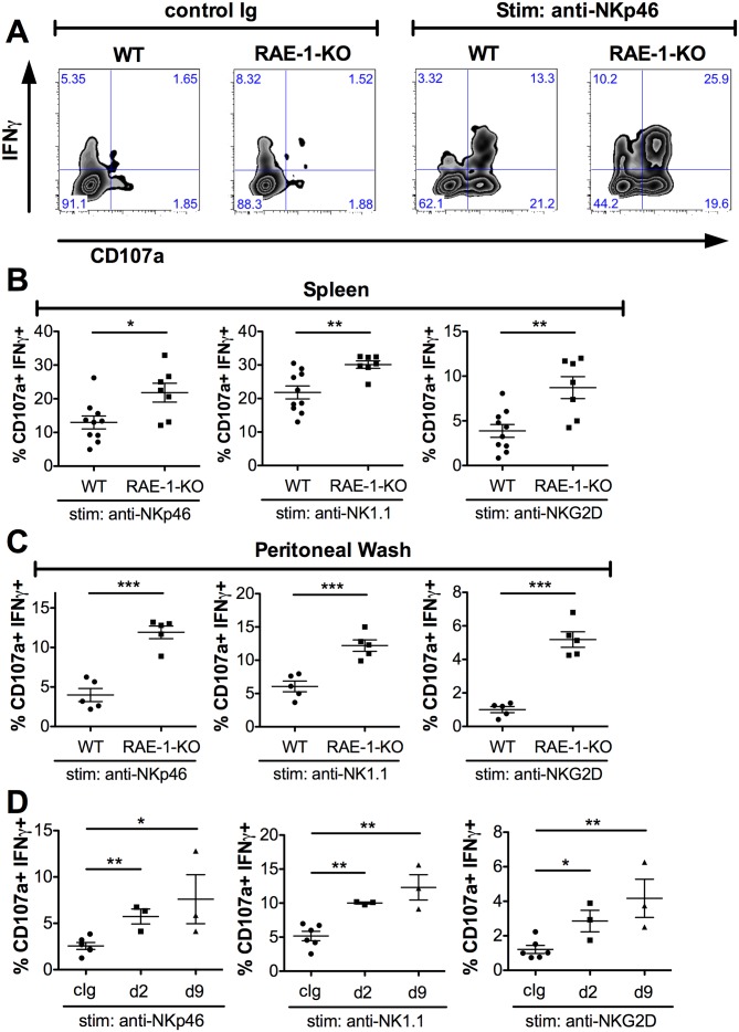
Figure 2. Endogenous RAE-1ε negatively regulates NK responsiveness.

(A) WT or RAE-1-KO splenic NK cell IFNγ production and degranulation (CD107a) after 5 hr ex vivo stimulation with platebound control Ig or anti-NKp46. (B and C) Percentage of activated (IFNγ- and CD107a-double-positive) splenic or peritoneal NK cells from WT or RAE-1-KO mice after ex vivo stimulation with the indicated plate-bound antibody. Data are representative of >4 independent experiments. (D) Percentage of activated peritoneal NK cells after ex vivo stimulation from mice given control Ig or anti-RAE-1ε for the indicated time. Data are representative of two independent experiments. Statistical significance was determined using two-tailed unpaired Student’s t tests. Data represent means ± SEM.

Figure 2.

Figure 2—figure supplement 1. Normal NK cell cellularity and differentiation but enhanced NK-mediated tumor cell killing in RAE-1-knockout mice.

Figure 2—figure supplement 1.

(A, left panel) NK cells from WT and RAE-1-KO mice as percentage of live splenocytes. Data are representative of >4 independent experiments. (A, right panel) CD27 and CD11b 
expression on splenic NK cells from WT and RAE-1-KO mice. Data are representative of three independent experiments. (B) NK cells from WT, RAE-1-KO, or NKG2D-KO mice (n = 3) were pre-activated with a single injection of 200 g Poly I:C. Two days later, peritoneal wash cells were harvested, pooled, and used as effector cells to kill YAC-1 cells in a standard 51Cr in vitro cytotoxicity assay, at the indicated effector: target ratios. Data are representative of two independent experiments. Statistical significance was determined using unpaired two-tailed Student’s t tests (A) or two-way ANOVA (B). Data represent means ± SEM (error bars are not visible because they are small).