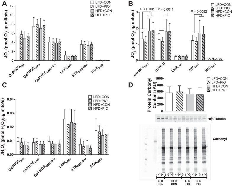
Fig. 4.
Skeletal muscle mitochondrial respiration and H2O2 emission. High-resolution respirometry was performed on mitochondria isolated from quadriceps muscle using glutamate-malate-succinate (GMS) or palmitoyl-l-carnitine for fatty acid oxidation (FAO) followed by inhibitors and uncoupler. Oxygen consumption (JO2) during glutamate-malate-succinate-based respiration was not different between diet or PIO groups when expressed relative to mitochondrial protein (mito) abundance (A). Oxygen consumption (JO2) during fatty acid oxidation (OxPHOSFAO) was increased with HFD with no effect of PIO when expressed relative to mitochondrial protein abundance (B). The addition of cytochrome c (cyto c) did not alter respiration and indicates that mitochondrial membranes were intact. There were no differences between groups in H2O2 emissions measured during glutamate-malate-succinate respiration (JH2O2; C). Oxidative damage was not different between diet or PIO treatment groups as measured by carbonyl modification to quadriceps muscle lysates (D). Representative images from blots for antibodies against oxidative damage and tubulin. Samples were analyzed for oxidative damage using nonderivatized (−) or derivatized samples (+) and were not boiled such that tubulin has an apparent molecular weight that is higher than predicted. Data were compared with two-way ANOVA (diet × drug). Data are means ± SD; n = 8–14 per group.

