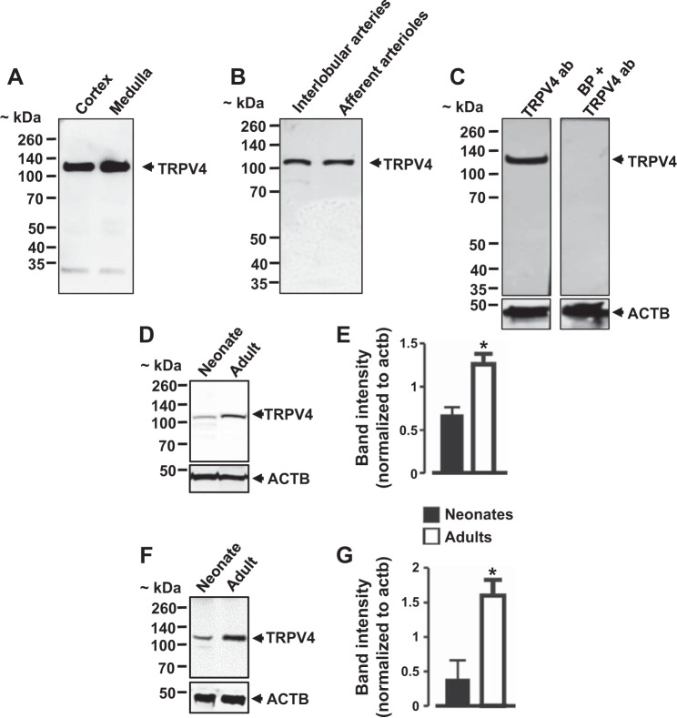
Fig. 2.
TRPV4 channel protein expression levels in porcine kidney and renal preglomerular vessels. A and B: Western blot images showing the expression of TRPV4 channels in neonatal pig renal cortex, medulla, and preglomerular vessels. C: a blocking peptide directed against TRPV4 antibody abolished TRPV4 immunoreactive band detection. Western blot images and bar graphs (n = 3 each) illustrating TRPV4 protein expression levels in neonatal and adult pig kidneys (D and E) and interlobular arteries (F and G). *P < 0.05 vs. neonates.

