Fig. 4.
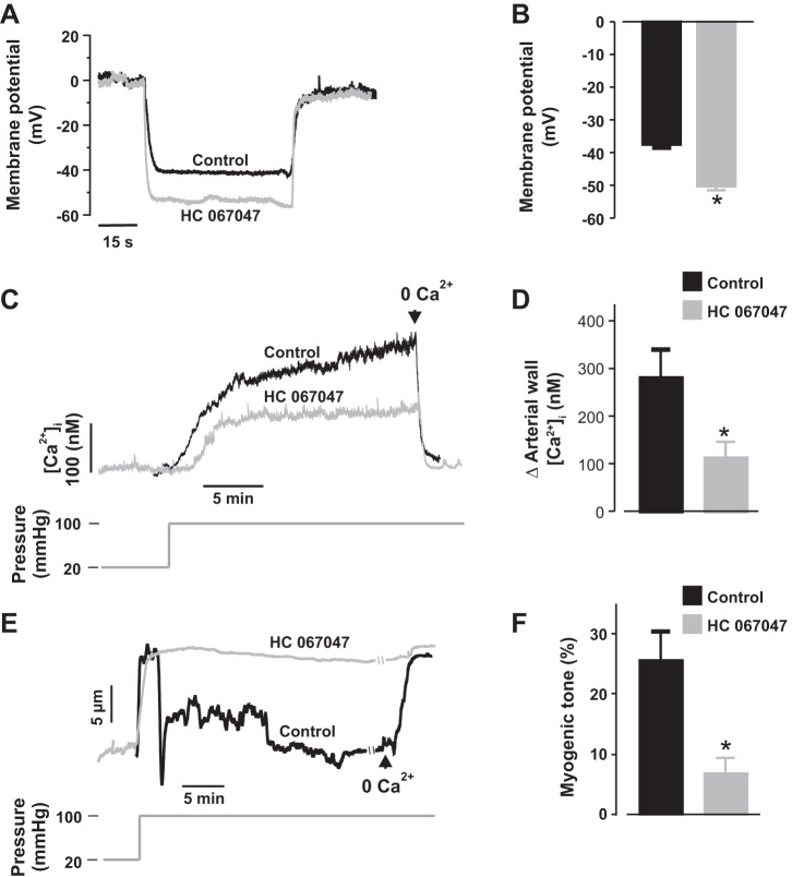
TRPV4 channels contribute to pressure-induced membrane depolarization, [Ca2+]i elevation, and constriction in neonatal pig renal preglomerular arteries. A and B: traces and bar graphs showing SMC membrane potentials in neonatal pig renal interlobular arteries that were pressurized to 100 mmHg in the presence of DMSO (control; n = 4) or HC 067047 (1 µM; n = 7). C and D: traces and bar graphs illustrating pressure-induced changes in [Ca2+]i concentration in neonatal pig renal interlobular arteries in the presence of DMSO (control; n = 4) or HC 067047 (1 µM; n = 5). E and F: traces and bar graphs showing myogenic tone in neonatal pig renal interlobular arteries that were pressurized to 100 mmHg in the presence of DMSO (control; n = 12) or HC 067047 (1 µM; n = 12). Vessels were pretreated and continuously superfused with DMSO or HC 067047. *P < 0.05 vs. control.

