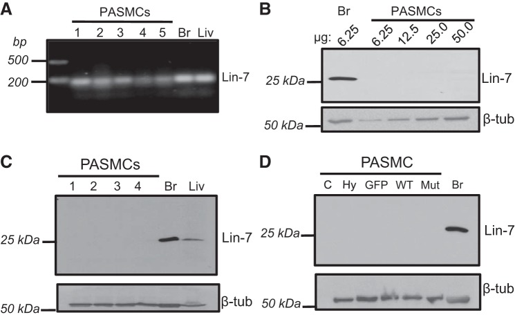
Fig. 8.
Lin-7 expression in rat pulmonary arterial smooth muscle cells (PASMCs). A: image showing PCR products for Lin-7A mRNA in PASMCs and control tissues, brain (Br) and liver (Liv). Each lane represents a sample from a different animal. B: representative immunoblot showing Lin-7 protein expression when increasing concentrations of total PASMC protein was loaded (6.25–50 µg) compared with brain (Br) lysate. C: representative immunoblot showing Lin-7 protein expression in PASMCs, Br and Liv and the corresponding β-tubulin (β-tub; housekeeping) for each sample. Each lane represents a sample from a different animal. For PASMCs, 50 µg of protein was loaded in each lane, compared with 6–10 µg for Br and Liv. D: immunoblot showing Lin-7 and β-tubulin protein expression in PASMCs under control conditions (C), exposed to hypoxia (Hy; 4% O2 for 24 h), or infected with viral constructs containing GFP (GFP), wild-type AQP1 (WT), or AQP1 lacking the COOH-terminal tail (Mut). Brain (Br) was used as a positive control. For PASMCs, 50 µg of total protein were loaded per lane, compared with 10 µg total protein for Br.

