Fig. 5.
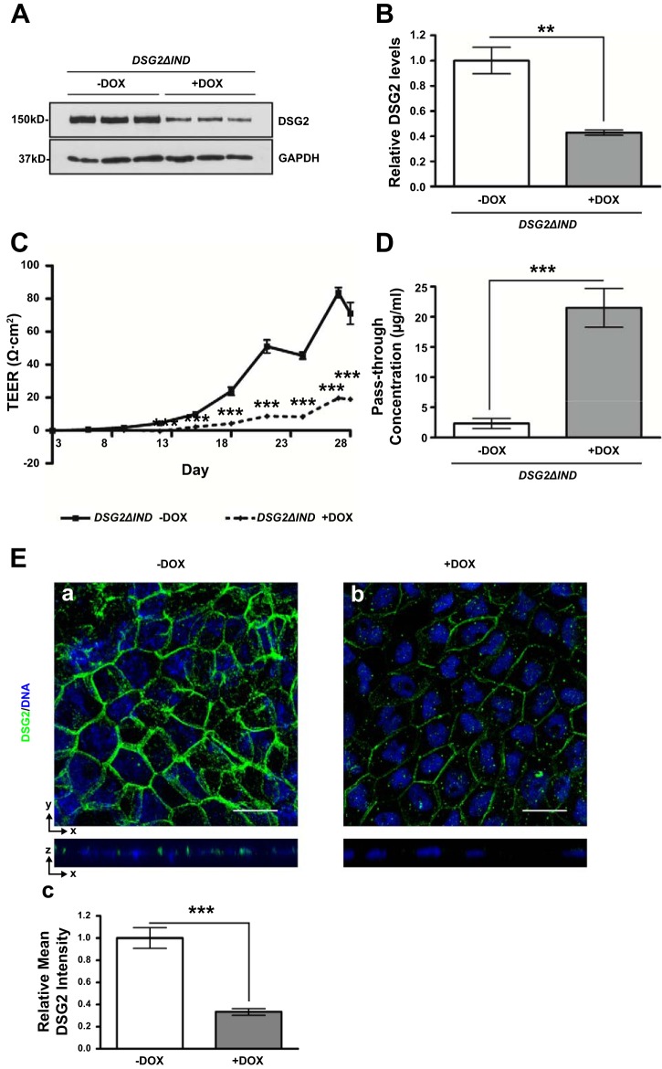
Epithelial barrier function is impaired in DSG2 knockdown Caco-2 BBe cells. Caco-2 BBe DSG2ΔIND cells were seeded in Transwell plates. On day 3 of the culture, cells were treated with water or doxycycline to induce expression of shRNA. Cells were maintained for a total of 28 days. A: Western blot analysis of the DSG2 protein levels in Caco-2 BBe DSG2ΔIND –DOX and Caco-2 BBe DSG2ΔIND +DOX cells (bottom) on day 28. A result from 3 independent experiments is shown. B: quantitative representation of DSG2 protein levels in 3 independent experiments, normalized to GAPDH. Data represent means ± SE (n = 3), **P < 0.01, Student’s t-test. C: transepithelial electrical resistances of Caco-2 BBe DSG2ΔIND monolayers grown in absence (–DOX, solid line) and presence (+DOX, broken line) of doxycycline were measured during 28 days of culture. Data represent means ± SE (n = 6), ***P < 0.001, Student’s t-test. D: permeability to FITC-4 kDa dextran measured on day 28. Data are shown as pass-through concentrations of FITC-dextran. Empty boxes, –DOX; filled boxes, +DOX. Data represent means ± SE (n = 6), ***P < 0.001 by Student’s t-test. E: immunofluorescence staining of DSG2 in Caco-2 BBe DSG2ΔIND –DOX and Caco-2 BBe DSG2ΔIND +DOX cells collected on day 28 of culture. The top part of each panel shows X–Y projections obtained from Z-stacks, and companion Z-plane cross sections are shown on bottom. DNA was counterstained with TO-PRO3. Images were taken at ×400 magnification. Scale bars indicate 20 µm. Relative mean fluorescence intensity was quantified (c). Data represent means ± SE (n = 6), ***P < 0.001 by Student’s t-test.

