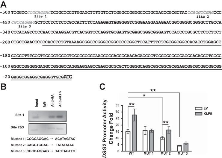
Fig. 7.
KLF5-binding sites are identified in DSG2 promoter. A: sequences of the potential binding sites for KLF5 are shown in gray and italic within 500 bp upstream of the start codon of DSG2, with transcript underlined. B: PCR products of DSG2 promoter sequences in input cell lysates and immunoprecipitations with rabbit IgG, rabbit HA antibody, and KLF5 antibody using HEK 293T cells cotransfected with pGL3BDSG2(500) and pMT3-HA-KLF5. Sequences of the site-directed mutation of the binding sites are shown. C: DSG2 promoter activity measured with luciferase assay. HEK 293T cells were transfected pEGFP-ΔEGFP or pEGFP-ΔEGFP-KLF5-HA together with pLightSwitch, pLightSwitch-DSG2(500), or pLightSwitch-DSG2(500) mutant promoter reporter plasmids. Data represent means ± SE (n = 18), *P < 0.05 and **P < 0.01 by Student’s t-test.

