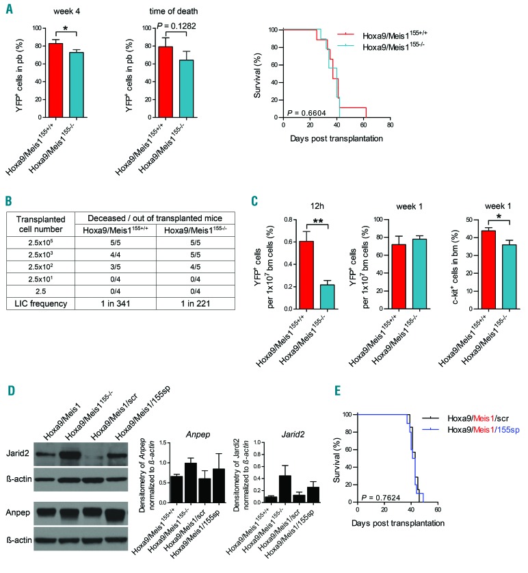
Figure 4.
Absence or depletion of miR-155 does not alter leukemogenicity of Hoxa9/Meis1 cells. (A) Engraftment in peripheral blood (PB) after 4 weeks (left panel), at the time of death (middle panel, n=7/arm) and survival (right panel) of mice transplanted in two independent experiments with two biological replicates of Hoxa9/Meis1155+/+ (n=9) or Hoxa9/Meis1155−/− (n=9) cells. Percentage of yellow fluorescent protein (YFP)+ cells was measured by flow cytometry in PB of mice. (B) Limiting dilution assay of Hoxa9/Meis1155+/+ and Hoxa9/Meis1155−/− transplanted cells with indicated numbers of mice transplanted for each arm. Leukemia initiating cell frequency was calculated using L-cal™. (C) Left: homing percentage of Hoxa9/Meis1155+/+ and Hoxa9/Meis1155−/− (n=5/arm) cells transplanted into non-irradiated recipient mice and assessed 12h post transplantation. Middle: percentage of Hoxa9/Meis1155+/+ and Hoxa9/Meis1155−/− (n=5/arm) cells transplanted into non-irradiated recipient mice and assessed one week post transplantation. Right: percentage of c-kit positive cells in Hoxa9/Meis1155+/+ and Hoxa9/Meis1155−/− (n=5/arm) cells transplanted into non-irradiated recipient mice and assessed one week post transplantation. (D) Left: western blot of Jarid2 and Anpep in Hoxa9/Meis1155+/+ and Hoxa9/Meis1155−/− as well as Hoxa9/Meis1/155 sponge and Hoxa9/Meis1/scr-transduced cells. Right: densitometry of Anpep and Jarid2 normalized to β-actin (gene/β-actin) using the ImageJ software (n=2 biological replicates). (E) Survival curves of two independent transplantation experiments, where 1000 cells of Hoxa9/Meis1/155 sponge (n=10) or Hoxa9/Meis1/scr, (n=7) cells were transplanted. See also Online Supplementary Figure S4 and Online Supplementary Table S7. BM: bone marrow; LIC: Leukemia-initiating cell.

