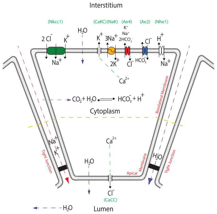
Fig. 1.
Schematic diagram of the salivary acinar cell model. We distinguish the basal and lateral sides (basolateral) to the apical side of the plasma membrane. Perfusion studies demonstrated different potentials on each portion of the acinar membrane (Young, 1968). In the diagram these are separated by a yellow line. The basolateral membrane portion contains Nkcc1 (green), NaK-ATPase (yellow), Ae4 (red), Ae2 (blue), Nhe1 (white), and Ca2+-activated K+ channels. The cell membrane is permeable to CO2; carbonic anhydrases in the cytoplasm catalyze the reaction of CO2 and water to form carbonic acid, which dissociates into and H+. The apical membrane contains a Ca2+-activated Cl− channel. Both apical and basolateral membranes are permeable to water. Finally, we have included paracellular K+ and Na+ currents along with a paracellular water flow. Although the apical membrane also contains Ca2+-activated K+ channels, these are omitted from the model for simplicity.

