
Figure 2.
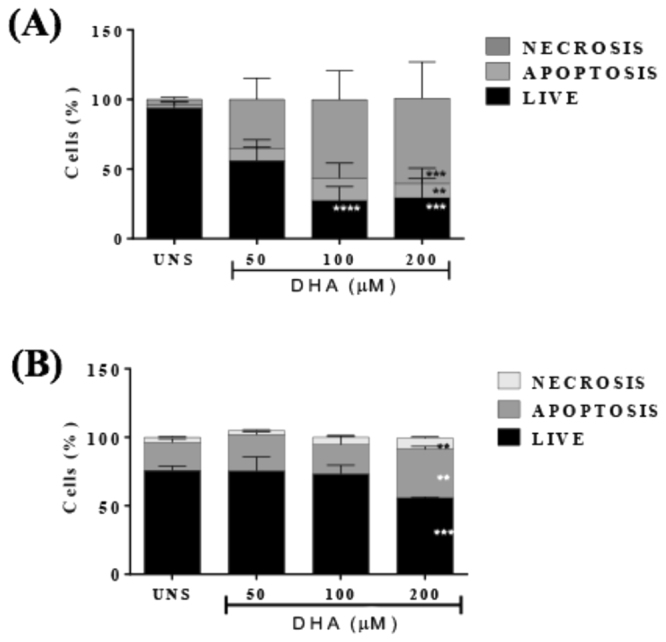
- DHA led to cell death in breast cancer cells. MDA-MB-231 (A) and 4T1 breast cells were stimulated with different concentrations of DHA for 24 h and cell death was assessed by labeling cells with annexin-V and propidium iodide followed by flow cytometry analysis. The percentage of necrotic, apoptotic or live cells is indicated. Data are representative of five independent experiments. UNS represents unstimulated cells. Each point represents the average percentage ± SD. (n = 5) *p < 0.05.