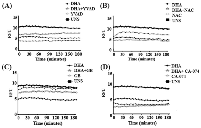
Figure 7.
–DHA induced membrane pore formation dependent on caspase-1 activation, lysosomal damage and ROS formation. MDA-MB-231 cells were pre-treated for 1 h with caspase-1 inhibitor Ac-YVAD-cmk 100 μM (A) or ROS inhibitor N-acetyl-cysteine NAC 5 mM (B), potassium efflux inhibitor glyburide GB 150 µM (C) and CA-074, a lysosomal cathepsin B inhibitor 50 µM (D). Then, cells were stimulated with 100 µM DHA for 180 minutes and propidium iodide uptake was measured by spectrophotometry. Graphics are representative of three independent experiments (n = 5 each). UNS: Unstimulated cells. RFU: Relative fluorescence units.

