Figure 3.
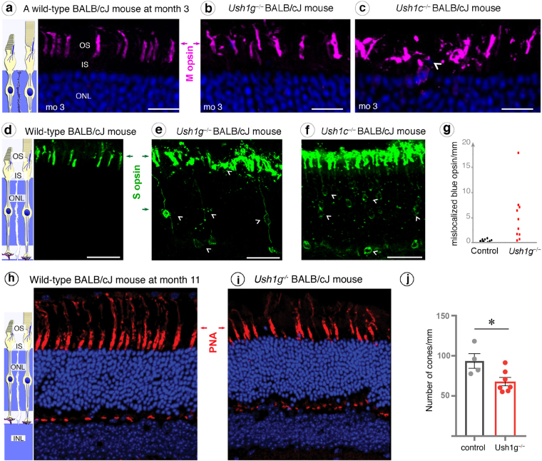
Mislocalization of cone opsins in Ush1c−/− and Ush1g−/− BALB/cJ mice and cone loss in Ush1g−/− BALB/cJ mice. (a–c) Immunolabeling of M opsin on retinal cross-sections at month 3 (mo 3), showing that the outer segments (OS) are normal in shape in wild-type (a) and Ush1g−/− BALB/cJ mice (b), whereas the outer segments in Ush1c−/− BALB/cJ mice are disorganized (c, white arrowhead). (d–g) The S opsin immunolabeling, which was restricted to the outer segment in wild-type BALB/cJ mice (d) revealed alterations to the outer segments in the two mutant mice, with mislocalized labeling extending over the entire photoreceptor cell body (white arrowheads, e and f). (g) Quantification of cone cells with mislocalized immunolabeling for S opsin (number of cells/mm retinal cross-section, p < 0.0001, Student’s t-test, n = 10 in each group). (h–j) Lectin PNA staining on retinal cross-sections in 11-month-old control (h) and Ush1g−/− (i) BALB/cJ mice showing a reduction in the number of PNA-stained cone photoreceptor inner/outer segments in Ush1g−/− (red) BALB/cJ mice (j, number of cells/mm retinal cross-section, p < 0.05 n = 4 control animals, n = 7 Ush1g−/− mice). The scale bars represent 5 µm in (a–c), 10 µm in (d–f) and 50 µm in (h and i). ONL: outer nuclear layer, INL: inner nuclear layer, IPL: inner plexiform layer.

