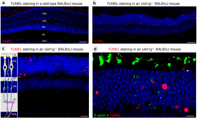
Figure 4.
Photoreceptor apoptosis in Ush1g−/− BALB/cJ mice. (a–d) TUNEL staining in wild-type (a) and Ush1g−/− (b–d) BALB/cJ mice at 3 mo. Apoptotic cells (labeled in red) are detected in the outer nuclear layer (ONL) of Ush1g−/− BALB/cJ retina (b–d), whereas such cells were completely absent from the retina of control BALB/cJ mice (a). Note the apposition of the TUNEL labeling and the mislocalized S opsin immunolabeling (white arrowhead, d). The scale bars represent 50 µm in (a and b), 20 µm in (c), and 15 µm (d). OS: outer segment, INL: inner nuclear layer, IPL: inner plexiform layer.

