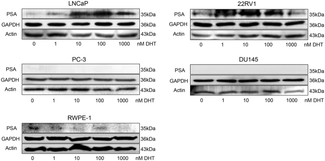
Figure 1.
Validation of DHT treatments by Western blot. AR+ cells (LNCaP and 22RV1), AR − cells (DU145 and PC-3), and normal prostate epithelial cell (RWPE-1) were cultured using RPMI 1640 medium containing 10% charcoal-stripped FBS for 24 hours, and then treated with different DHT treatments. Validation of DHT treatments were performed through Western blot experiments using PSA as a marker, and GAPDH and Actin as control proteins. The grouping of blots were cropped from different parts of the same gel, or from different gels. Full-length unadjusted Western blot images for this figure as shown in Supplementary Fig. S3.

