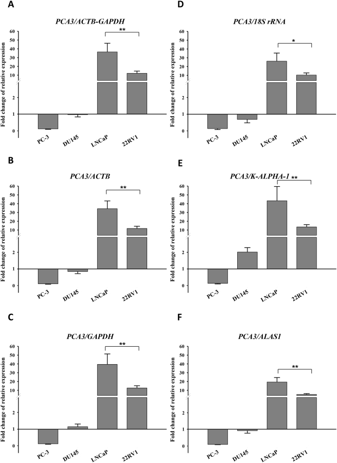
Figure 6.
Relative quantification of PCA3 expression depends on mRNA reference gene. Cells were cultured using RPMI 1640 medium containing 10% charcoal-stripped FBS for 24 hours prior to treatment with different DHT concentrations. The cDNA was prepared from these cells for the amplification of PCA3 and mRNA reference genes by qPCR. Relative expression of PCA3 across all cell lines was normalised by the best reference genes combination (ACTB-GAPDH) (A), by the most stable single gene ACTB (B) or GAPDH (C), by the least stable single gene 18 S rRNA (D), or K-ALPHA-1 (E), or ALAS1 (F). Y-axis indicates the fold change of relative expression of PCA3 in RWPE-1 cells set to 1. Error bars indicate the standard error (±SE) evaluated from three biological replicates. * and **Indicate P < 0.05 and P < 0.01, respectively.

