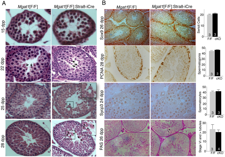
Figure 1.
Onset of morphological changes in Mgat1[F/F]:Stra8-iCre testes. (A) Representative control Mgat1[F/F] and Mgat1[F/F]:Stra8-iCre testis sections stained with hematoxylin and eosin at 15–28 dpp (n = 3–4 mice per genotype; 20–30 tubules observed per section). At 22 dpp round spermatids were present in control and Mgat1 cKO tubules (arrows). At 25 dpp, few elongated spermatids were seen in Mgat1 cKO tubules and some MNC were observed in a few tubules (asterisks, inset). At 28 dpp, Mgat1 cKO tubules contained MNC comprised of fused spermatids (asterisks, inset and Supplementary Fig. S1). Images were scanned at 40× in a Perkin Elmer Scanner. (B) Sertoli cells were identified by anti-Sox9 Ab, spermatogonia by anti-PCNA Ab; primary spermatocytes by anti-Sycp3, and acrosomes by PAS. Histograms show numbers of stained cells per 20–30 tubules per section, and numbers of mice examined. Tubules at stage VI and beyond based on morphology with PAS+ acrosomes were counted per 50 tubules. Histograms represent mean ± SEM. Images were photographed at 20×. Controls for antibody specificity are shown in Supplementary Fig. S2.

