Figure 1.
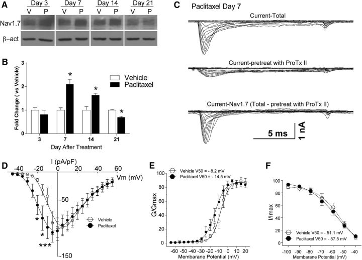
Increased expression and function of Nav1.7 in DRG neurons in rats with paclitaxel-induced peripheral neuropathy. A, The representative Western blot images demonstrate that the expression of Nav1.7 was increased in DRGs by day 7 of chemotherapy through day 14, whereas the expression of Nav1.7 then fell below the baseline level at day 21. The bar graphs in B summarize the grouped data and indicate that the level of expression of Nav1.7 in the dorsal root ganglia in the paclitaxel-treated rats (black bars) was significantly higher than in the vehicle-treated rats (white bars). C, Representative voltage-clamp recordings of Nav1.7 current in acute dissociated DRG neurons of day 7 paclitaxel-treated rats collected using a subtraction protocol with the administration of Nav1.7-selective blocker ProTx II, as described in Results, the mean current density–voltage relationships are plotted in D. Shown in E is a comparison of the voltage dependence of activation for Nav1.7 channels in vehicle- and paclitaxel-treated animals. In F, the comparison of steady-state inactivation for Nav1.7 channels is shown. β-act, β-actin; V, vehicle; P, paclitaxel. *p < 0.05, ***p < 0.001.

