
Figure 3.
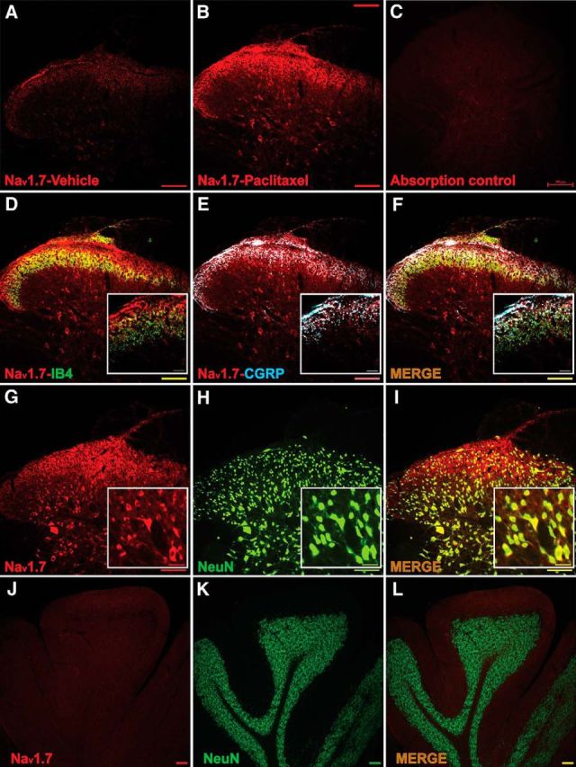
The expression of Nav1.7 is increased and colocalized with IB4, CGRP, and NeuN in spinal cord segments L4–L5 at day 7 after treatment with paclitaxel. A, B, Representative immunohistochemical (IHC) images show that expression of Nav1.7 (red) in spinal cord was quite pronounced compared with vehicle (A) by day 7 after treatment with paclitaxel (B). In C, the absorption control shows no staining. D, E, Double IHC shows that Nav1.7 (red) was expressed in some IB4-positive (D, green, with colocalization shown in yellow) and especially were expressed in CGRP-positive (blue) neuron terminals (E, colocalization indicated in purple). In F, the merged image indicates that Nav1.7 expression was upregulated in a large percentage of peripheral afferent terminals positive for CGRP. G–I show that Nav1.7 was expressed in NeuN-positive spinal cells in deeper lamina following paclitaxel treatment. J–L are a further test of antibody specificity as no expression of Nav1.7 was found in the cerebellum. Scale bars: lower-scale images, 200 μm; higher-magnification images, 50 μm.