Figure 1.
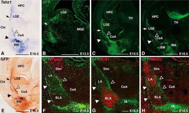
Tshz1GFP drives GFP in the LGE and ITC clusters. A, In situ hybridization showing Tshz1 gene expression in the LGE, LMS (arrow), and ITC clusters. Solid arrowhead indicates lateral. Open arrowhead indicates medial. B, Immunohistology for GFP (green) in E13.5 Tshz1GFP/+ mice shows GFP protein extending from the dLGE SVZ to the mantle zone. C, D, At E15.5 (C) and E18.5 (D), GFP protein expression refines to a distinct stream (arrow) emerging from the dLGE and several contiguous clusters in the amygdala comprising the lateral paracapsular clusters (solid arrowheads), medial paracapsular clusters (open arrowheads), and IA. E, GFP immunohistochemical staining recapitulates the Tshz1 expression pattern (A). F, Amygdalar GFP staining colocalizes with the ITC marker Foxp2. G, H, GFP-labeled cells in the amygdala surround cells expressing the BLA marker Er81 (G) and the LA marker Mef2c (H). BM, Basomedial amygdala; Ctx, cortex; HPC, hippocampus; LGE, lateral ganglionic eminence; MA, medial amygdala; MGE, medial ganglionic eminence; Stm, striatum; TH, thalamus. Scale bars: A–E, 500 μm; F–H, 100 μm.

