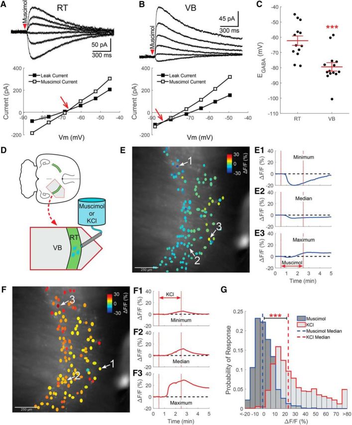
Figure 2.

RT neurons maintain a relatively low [Cl−]i. Gramicidin perforated patch recordings of muscimol-induced (100 μm, 10 ms) currents from RT (A) and VB (B) neurons at various command potentials. The leak current has been subtracted from the representative traces. The intersection of the prestimulation leak current and the muscimol-induced current was used to determine the GABAA receptor-mediated equilibrium potential (EGABA, red arrow). C, The EGABA of RT neurons was more depolarized than in VB neurons, yet remained at levels that likely support inhibitory GABAergic signaling. D, GCaMP6s was expressed in RT neurons for calcium imaging experiments and a local perfusion system provided timed delivery of muscimol (5 μm) or elevated KCl (+10 mm) to the imaged RT nucleus. E, Two minute application of muscimol mostly decreased the fluorescence of RT neurons. ROIs drawn around GCaMP6s-expressing RT neurons are colored according to their peak change in fluorescence. Representative examples of neurons displaying the minimum (E1), median (E2), and maximum (E3) fluorescence change in a particular brain slice following muscimol application. F, Two minutes of elevated KCl produced a nearly uniform increase in the fluorescence of RT neurons. Examples of the minimum (F1), median (F2), and maximum (F3) fluorescence changes evoked by elevated KCl application. G, Histograms comparing induced responses in all cells imaged, across multiple animals, shows that the median response (dotted line) to muscimol was a slight decrease in fluorescence. In contrast, mild depolarization with elevated KCl produced a robust increase in fluorescence. Bin size: 5ΔF/F(%). ***p < 0.001.
