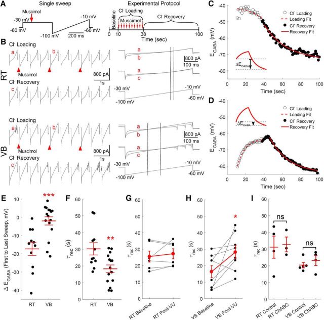
Figure 7.
Recovery from Cl− loading is limited in RT neurons. A, Schematics of protocol for Cl− loading experiments. Neurons were voltage-clamped at −30 mV for 500 ms and then ramped from −100 to −10 mV over a duration of 500 ms (left). This protocol lasted one second and was repeated 100 times per cell (right). After 10 s of baseline recording, Cl− was loaded by applying 10 puffs of muscimol (20 ms, 100 μm), once every 3 s, while the neuron was held at −30 mV. B, Gramicidin perforated patch recordings of RT and VB neurons during measurement of Cl− loading and recovery. EGABA was measured by finding the point in the voltage ramp where the membrane current responses from before (a) and during (b) muscimol application intersected (marked by dotted line). Cl− recovery was measured by tracking the change in EGABA following the last application of muscimol (a vs c). Single-exponential functions were fit to the shifting EGABA occurring in RT (C) and VB (D) neurons to determine time constants for Cl− loading and recovery. E, The change in EGABA (ΔEGABA) was measured between the start of Cl− loading and the final reading during Cl− recovery (C, inset). This measurement indicates that chloride loading occurs rapidly in RT neurons. F, The basal Cl− recovery rate (τrec) was slower in RT than in VB neurons. The τrec in RT neurons was unaffected by a 10 min application of VU0463271 (G, 10 μm), but became slower in VB neurons (H). I, A 2 h incubation in ChABC (0.4 U/ml, 37°C) did not alter the τrec of either RT or VB neurons. *p < 0.05, **p < 0.01, ***p < 0.001.

