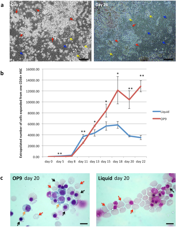
Figure 1.
OP9 co-culture delays differentiation of erythroid cells. CD34+ cells were cultured with and without OP9 stromal cells in an erythroid differentiation culture. (a) Erythroblasts (red arrows) attached to OP9 stromal cells (blue arrows) at day 7 and day 26 of the co-culture taken under phase contrast microscopy (scale bars 100 μm). Yellow arrows indicate adipocytes. (b) Comparison of the number of erythroid cells from day 0 to day 22 in control liquid and OP9 co-culture (for OP9 co-culture, only detached cells being counted) (mean ± SD; n = 3, *p < 0.05, **p < 0.0; student’s t test). (c) Erythroid cells from OP9 co-culture and liquid culture at day 20 stained with Leishman reagent and analyzed by light microscopy (scale bars 10 μm). Orange arrows indicate polychromatic erythroblasts, black arrows indicate orthochromatic erythroblast, red arrows indicate reticulocytes.

