Figure 1.
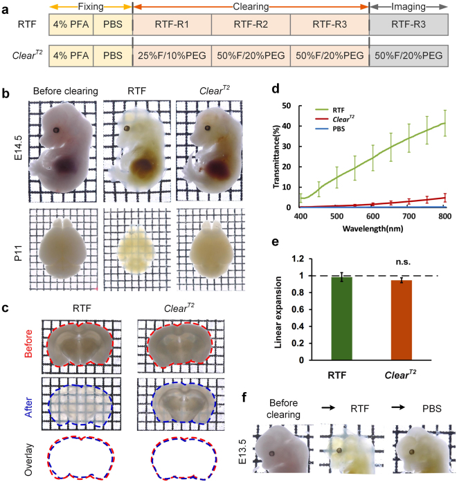
Rapid optical clearing using RTF. (a) Clearing steps of RTF and ClearT2. The boxes in the diagram only indicate the step. (b) Whole embryos (E14.5) and neonatal (postnatal day 11, P11) whole-brain samples cleared with RTF and ClearT2 overnight (transmission images). Grid size, 1.45 mm × 1.45 mm. (c) Adult brain slices (1-mm-thick) cleared with various clearing protocols. The outlines of the brain slices were drawn with dashed lines. Grid size, 1.45 mm × 1.45 mm. (d) Transmittance curves of the cleared mouse brain sections (1-mm-thick) (mean ± s.d., n = 6). (e) Normalized linear expansion of adult brain slices (1-mm-thick) after optical clearing (mean ± s.d., n = 6). Mann-Whitney U test was used to compare the difference of ClearT2 with RTF. n.s., not significant. (f) Clearing of RTF is reversible with PBS. Grid size, 1.45 mm × 1.45 mm.

